This may be the cause of melanin spots
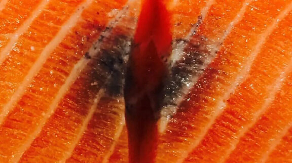
Melanin spots in salmon fillets have long been a major quality problem and lead to significant economic losses. Researchers now believe that the development of the spots is linked to changes in the fatty tissue.
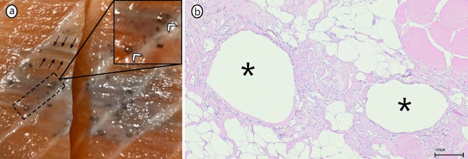
Research into melanin spots has been carried out for over 20 years at the Veterinary College at the Norwegian University of Life Sciences (NMBU), and scientists are now facing a significant breakthrough. In a recently published peer-reviewed article, developmental stages of melanin spots are described in line with “fat necrosis”, a condition that can also occur in other animals and humans, where the body’s immune system reacts against dying fat cells and their contents.
In humans, fat necrosis can occur in tissue that contains a lot of fat, such as under the peritoneum, in breast tissue, and in the subcutaneous tissue. The salmon stores significant amounts of fat in the muscles, and therefore fat necrosis can occur here. In addition, both bleeding and melanin spots occur mainly in the front areas of the fillet, which are also the fattest.

“Previously, we mainly focused on the muscle cells, but now we have gone in depth on the fat cells. We started by mapping the fat’s distribution in the muscles, as a basis for understanding the changes in the spots,” says associate professor Håvard Bjørgen.
Bleed in fat tissue
He explains that researchers see consistency with the development of fat necrosis in humans, where the condition begins as a bleed in the fat tissue, with subsequent tissue hypoxia and cell death.
“Dying fat cells release fat that can collect cysts or form so-called granulomas, which are chronic accumulations of inflammatory cells. Both parts are possible outcomes. The point is that the fish’s immune system is unable to handle the enormous amounts of free fat, and thus the fish gets chronic changes in the muscles.”
As of now, the researchers cannot say which characteristics of the fat are decisive for the development of fat necrosis.

Vegetable oils
“We consider it most likely that a high content of vegetable oils in the feed gives a fat composition with too much polyunsaturated fatty acid and correspondingly too little saturated fat. This is a known cause of fat necrosis in other animals. In addition, an unfavourable relationship between the fatty acids omega-6 and omega-3 can act as fuel on the fire,” says Professor Erling Olaf Koppang.
Polyunsaturated fatty acids are prone to oxidation; a process where free radicals “attack” the fatty acids and cause cell membrane damage.

“The pigment melanin is a powerful antioxidant. We see that the melanin is mainly around fat accumulations in the spots, and the melanin probably has a protective effect on the tissue that is exposed to oxidation,” says Koppang.
The subject group for anatomy at the Veterinary College,
NMBU has had projects on melanin spots more or less continuously since 2012 and
has throughout much of this period been funded by FHF - the Norwegian Fisheries
and Aquaculture Industry’s research funding body.
It's up to the industry
“Now it is up to the industry to put this knowledge to use and implement measures to avoid the problem of dark spots,” says Sven Martin Jørgensen, head of fish health at FHF.
“It is natural to look at the content of the feed recipes, and there are already farming companies that, for example, have increased the omega-3 content in the feed with positive effects, but you should probably also look at the effects of reducing the fat content overall,” concludes Jørgensen .
“The findings we now have show how important such long-term financial support is. It has provided continuity in our research, which has produced these important results,” says Koppang.
For more information, read Håvard Bjørgen's article, Deciphering the pathogenesis of melanized focal changes in the white skeletal muscle of farmed Atlantic salmon (Salmo salar), published in the Journal of Fish Diseases.


